

It is the science that deals with discomfort in the gum, connective tissue and bone around the tooth. Periodontological treatment; This includes early treatment of gum disease, removal of plaque and calculus on the teeth and ensuring a smooth root surface.
This process; It provides removal of bacteria and irritants that cause inflammation in the gums that may not require early treatment, surgical treatment. Mass gum loss can be prevented by treating gum diseases that manifest themselves in the clinic with gingival bleeding and gaping, and by informing the patient about this issue.
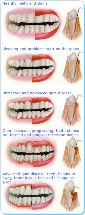
Healthy Gums

- It should be light pink in orange peel image;
- There should be a uniform gingival border that wraps the teeth at the root beginnings and follows their contours;
- There should be no redness, bloating or infection;
- Should not bleed with normal brushing and thread use;
- There should be no discomfort;
- It is solid and hard looking.
Diseased Gums
- Bleeding spontaneously or when using brush-floss,
- Redness, swelling and irregular appearance in the gums,
- Mild pain when pressing on the gums, inflammation leaking from the gums and / or tenderness in the tooth in that area,
- Withdrawal in gums and tenderness on exposed root surfaces,
- Black areas at the edges of the gums caused by tooth stones,
- Shaking, elongation and opening between the teeth,
- When you close your mouth, the feeling of change in your closing,
- Smell and bad taste due to inflammation,
- Itching sensation (in the gums)
- Constant bad breath
- Gums that can be easily separated from the teeth, move away and withdrawn
- Inflammatory discharge between teeth and gums
- Change in partial denture fit, deterioration

However, periodontal diseases can reach advanced stages without any findings. Therefore, it is extremely important to go to the dentist at regular intervals (6 months).
Bacteria Plate
The main cause of gum disease is a sticky transparent layer that clings to the tooth called bacterial plaque. You can notice the plaque by engraving on your tooth with your nail. One milligram of the plate contains between 200 and 500 million bacteria.
In addition, the following factors affect your gum health.
- SMOKING
- GENETIC FACTORS
- HORMONAL CHANGES
- Stress
- DRUG USE
- TEETHING OR CLEANING
- DIABETES-SUGAR DISEASE
- BAD NUTRITION
- BAD BROAD BRIDGE AND FILLINGS MADE
- ALLERGY DEVELOPING AGAINST KURON BRIDGE MATERIALS

Gum Disease Types
INGIVITIS
It is the simplest form of gum disease. It causes the gum to become red and swollen. The gums bleed quickly. Usually, at this stage, the patient does not experience much discomfort. With professional treatment and good care, gum health can be restored. If your oral care habits are insufficient, gingivitis starts moving towards periodontitis.
AGGRESSIVE PERIODONTITIS
In this type, it seems that there is no problem at first glance. On the contrary, the disease progresses aggressively and destructively. It may not respond to gum treatment and may need to be supported by antimicrobial (antibiotic) treatment.
CHRONIC PERIODONTITIS
There is progressive attachment (tissue between tooth and gum) and bone loss as a result of inflammation in the supporting tissues of the tooth. It is characteristic with pockets and withdrawal in the gums. It is the most common periodontitis variety. Although it is mostly seen in adults, it can be at any age. Attachment loss is usually slow, but rapid feed times can also occur.
PERIODONTITIS AS AN INDICATOR OF A SYSTEMIC DISEASE like diabetes….
NECROTIZING PERIODONTITIS
Inflamed necrotizing character in alveolar (bone tissue surrounding the tooth) bone, periodontal ligament (fibers between tooth and bone) and gum tissue. It is more often observed in people with an immune system suppression, malnutrition, and people with the AIDS virus.
Gingival Treatments:
In the treatment of gum disease, the depth of the pockets formed between the tooth and the gingiva needs to be measured with a special tool. Diagnosis is made according to the amount and depth of these pockets and treatment is planned. Since deep pockets will prepare a suitable environment for the rapid progression of gum disease, the purpose of the treatment is to make them as shallow as possible. Because it is impossible to completely clean the microorganisms that settle in the deep pockets by brushing and using dental floss.
DENTAL CLEANING AND ROOT SURFACE STRAIGHTENING (CURETAGE)
It is the most common and protective method of treating gum disease. In tartar cleaning, tartar and also called tartar are removed. Plaque is a sticky substance and the majority consists of bacteria. With the hardening of the plaque over time, dental stones are formed. Plaque and calculus attach to the tooth surface, especially to the root surface located below the gingival border. The root surfaces need to be leveled, as the plate tends to hold onto the rough surface. In this process, all dental stones are cleaned and irregularities on the root surface are removed.
In the early stages of the disease, this treatment is sufficient to control the condition in gingivitis. However, in advanced cases this may be the first step of treatment.
As this procedure may cause discomfort in some patients, local anesthesia may be applied to the area to be studied in these patients.
Ultrasonic tools and hand tools are used for scaling and smoothing the root surface. Ultrasonic instruments operate with air pressure or electricity. It has two components. The first is a relatively unsharp metal tip. This tip vibrates at high frequency and removes plaque and tooth stones from the surface. The second component is the water washing system which helps the metal tip to cool while it is working and to remove the residue from the tooth. The hand tools have sharp edges and these edges are leaned against the tooth and the stones on the tooth surface are removed. These hand tools are called scaler and curette. They are of various shapes and sizes for different teeth and different surfaces of the same tooth, and are not motor tools.
First of all, large plaque and tooth stones are removed from the crown and root surface of the tooth with ultrasonic tools. hand tools are used to remove all remaining material and straighten the root surfaces. While the dentist is working under the gingiva border, he cannot see plaque or dental stones. However, the roughness of the root surface directs it.
There may be hot-cold sensitivity and mild pain, which will disappear two to three days after treatment. Pain relievers can be used to eliminate pain.
GINGIVECTOMY AND GINGIVOPLASTY:
Gingivectomy is the surgical removal of gingival tissue. Gingivoplasty is the shaping of healthy gingival tissues around the teeth.
Gingivectomy is a treatment developed for the treatment of gum disease. Today it is also used for aesthetic arrangements.
There are two reasons for gingival tissue removal. The first reason is the presence of gingival pockets formed between the tooth and the gums. With the accumulation of food residues and bacterial colonies in these areas, cleaning difficulties arise. If these pockets contain gingival tissue only, they can be removed by gingivectomy.
In some cases, there may be a large amount of gums around the tooth. This prevents the teeth and gums from being kept clean. It is also a cosmetic problem. In severe cases, it may be sized to affect chewing and speech. As with epilepsy drugs, some medications can cause excessive gingival enlargement. Sometimes there may be no obvious reason.
The gums that are reshaped with the help of gingivoplasty have a more natural appearance. Disfigured or asymmetrical gums can be corrected with this procedure due to genetics, disease or trauma.
This process can also be done only due to cosmetic needs. Gingivoplasty is usually performed alone. However, in some cases, it can be applied after gingivectomy or graft application at the gingival border.
Before removing gingivectomy and gingivoplasty, dental calculus cleaning and root surface flattening may be required to remove dental calculus from existing gingival pockets.
Gingivectomy and gingivoplasties are usually done with hand tools. But it can also be done with electrosurgical instruments, laser and / or rotary instruments.
Local anesthesia is performed to numb the gums.
During the healing period, the mouth should be kept clean.
FLAP OPERATION
If periodontal pockets cannot be shallow by curettage and root surface straightening, the gum that creates the pocket is removed surgically.
The purpose of gingival flap surgery is to treat gum disease (periodontitis). This procedure is recommended for people with moderate or advanced gum disease. If gingival infection cannot be eliminated with a non-surgical method, gingival flap surgery is used. Gingival flap surgery can also be performed in conjunction with another procedure, bone surgery.
The region is primarily anesthetized with local anesthesia. The gums are separated from the teeth with the help of a hand tool. Thus, the periodontist can directly access the roots and the bone tissue supporting the tooth. Infected tissues are then removed between the teeth and holes (defects) in the bone. dental cleaning and root surface smoothing are applied. If there are defects in the bone, they can be reshaped by reshaping. During this process, bone corners can be softened using Er: YAG laser or rotary tools.
After these procedures, the gums are placed back on the teeth and fixed with stitches.
It is very important to keep this area as clean as possible while the surgical area is healing. You should continue brushing your teeth and flossing in the remaining areas.
If the periodontal bandage was used to protect the surgical area, it should be ensured that the plaque is removed gently from the teeth. Antimicrobial mouthwashes with chlorhexidine are usually prescribed after periodontal surgery. These mouthwashes do not remove the plaque, but it helps kill the bacteria and heal the mouth.
CROWN LENGTH OPERATION
It is a simple procedure to lengthen the parts of the teeth (visible in the mouth) above the level of the gums.
In periodontal surgical procedures, Soft Tissue Lasers are used successfully as well as classical surgical methods and effective results are obtained.
TISSUE GRAINS
Soft tissue grafts: It is the process of covering the apparent gingival recessions occurring in one or more teeth due to anatomical errors or gum diseases with a piece of soft tissue taken from a different part of the mouth (usually palate).
Hard tissue grafts: During flap operations, bone making is stimulated with artificial or natural bone bone applications in areas where bone loss is advanced.